Didactic Illustration
Show Me the Unseeable
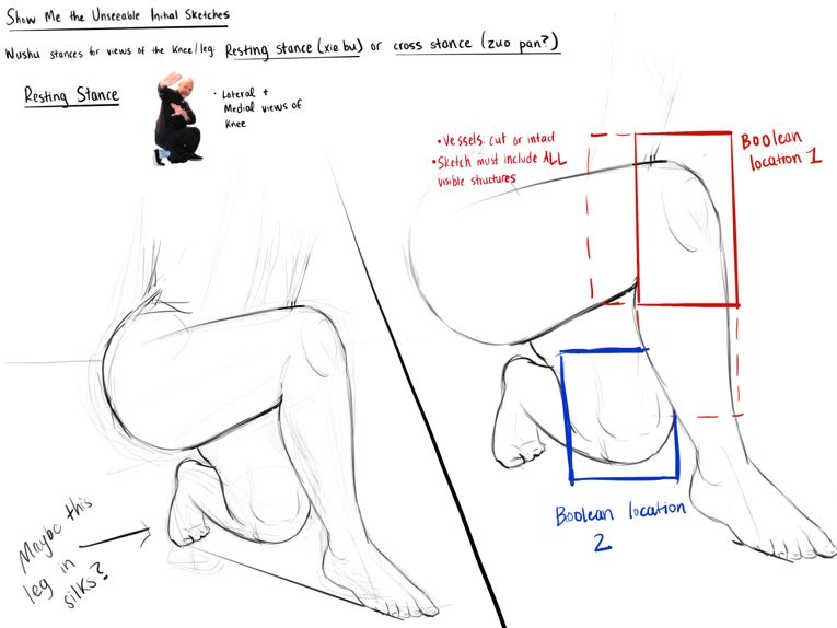
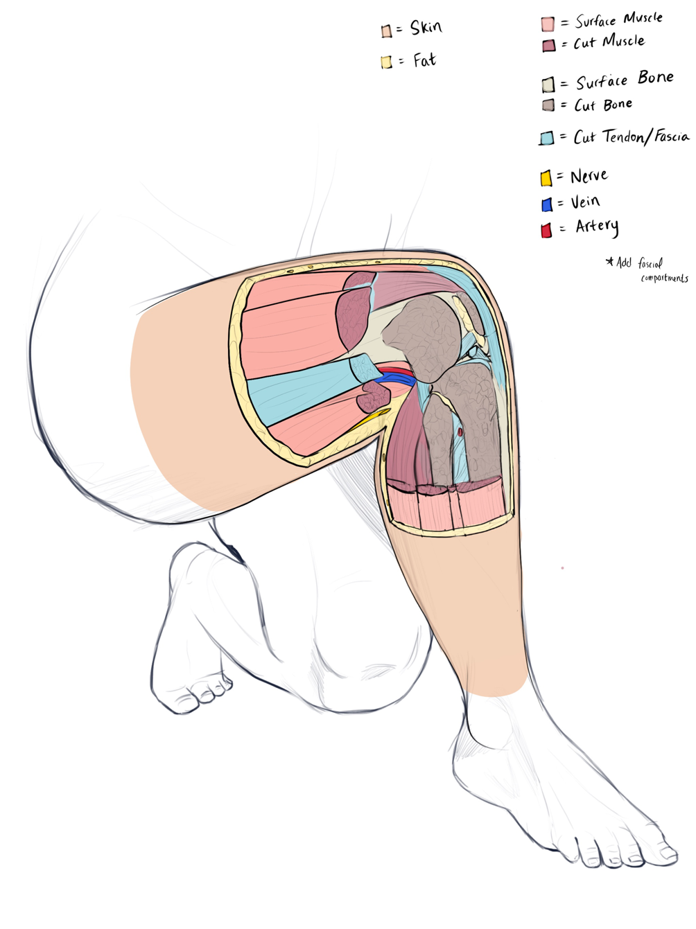
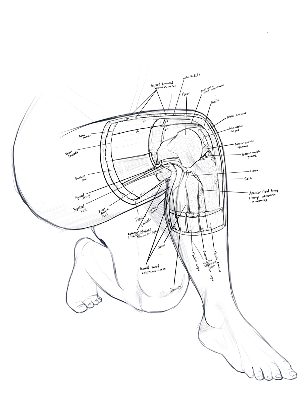
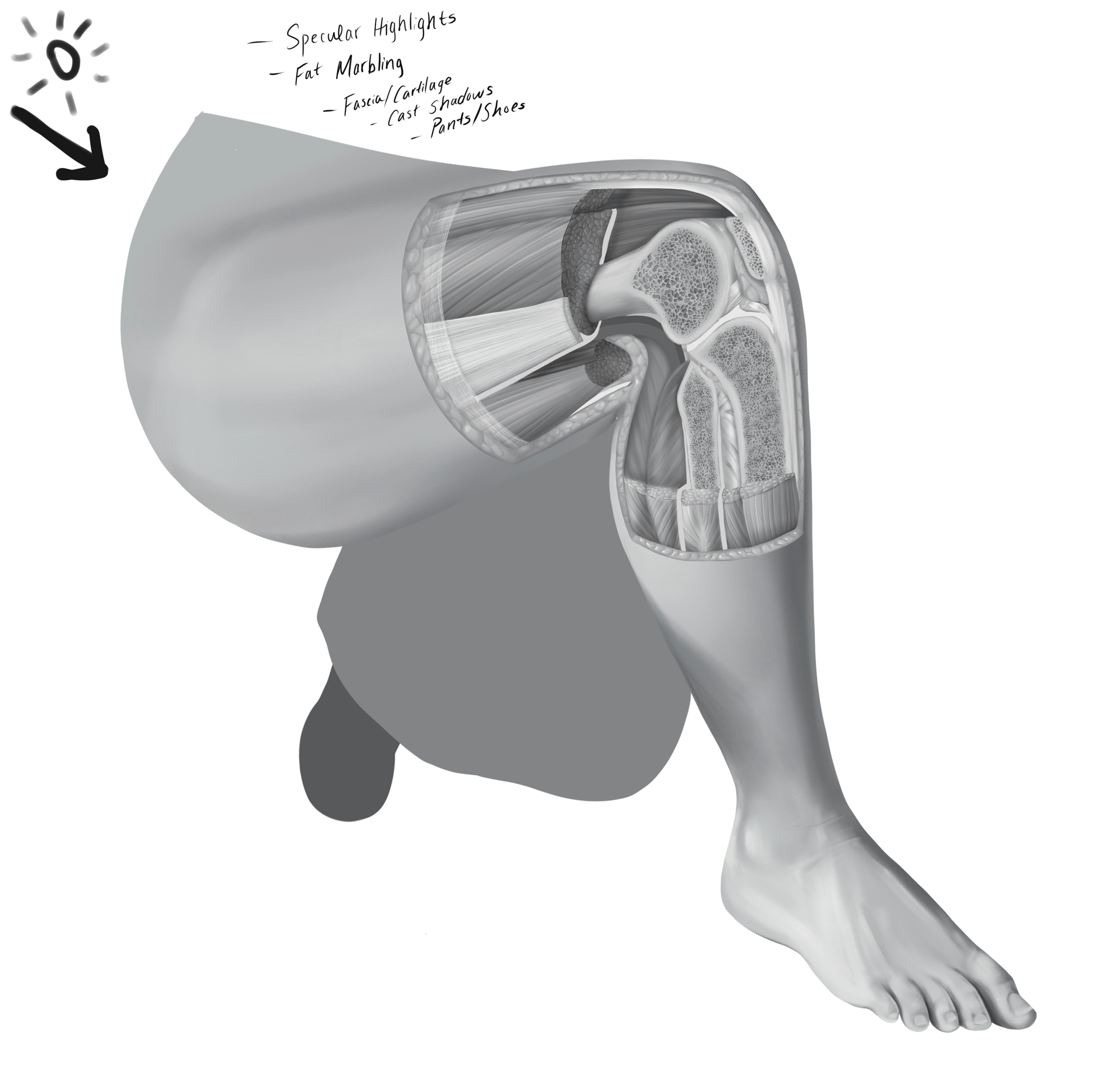
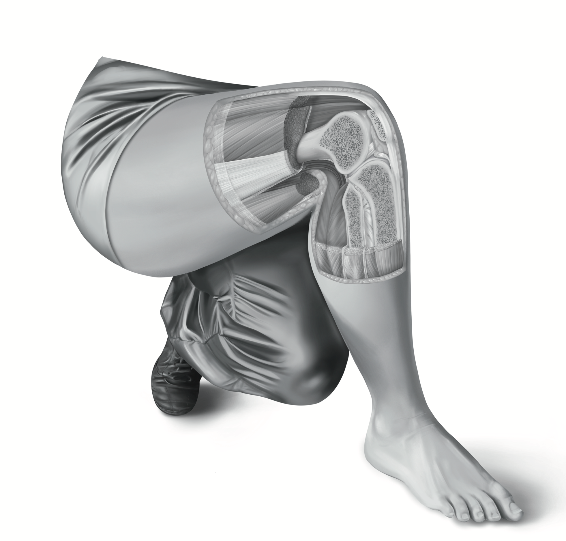
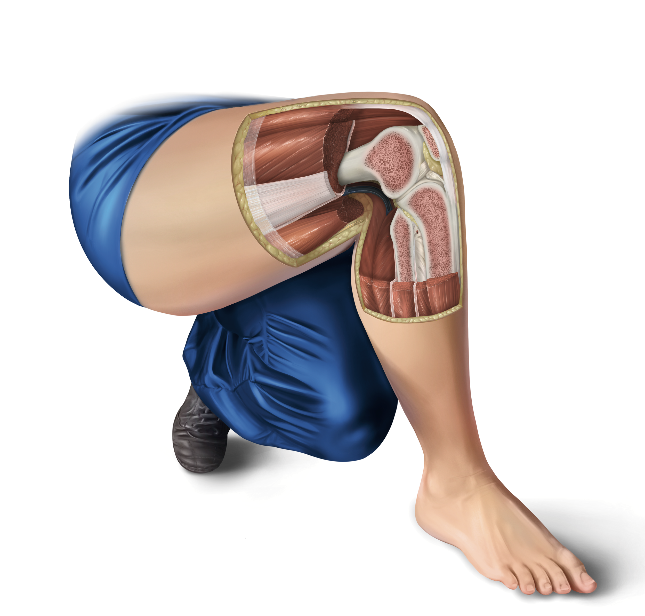
This painting reveals a previously “unseeable” Boolean cut of the human body. I selected this pose to demonstrate anatomy at work and draw upon my personal passion for martial arts.
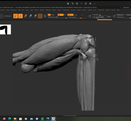
The illustration required meticulous anatomical knowledge, spatial reasoning, and knowledge of various rendering techniques. I constructed a 3D maquette for reference using Cinema4D and ZBrush. University of Toronto anatomist Dr. Anne Agur reviewed and approved the final illustration.
Inferior View of the Skull (with Mandible)
The carbon dust technique is a rite of passage for every medical illustrator. It provides unparalleled contrast and allows for meticulous detail, but requires painstaking precision from the artist.
This skull illustration was drawn from observation, reviewed for accuracy by University of Toronto faculty, and then rendered with the traditional medium.
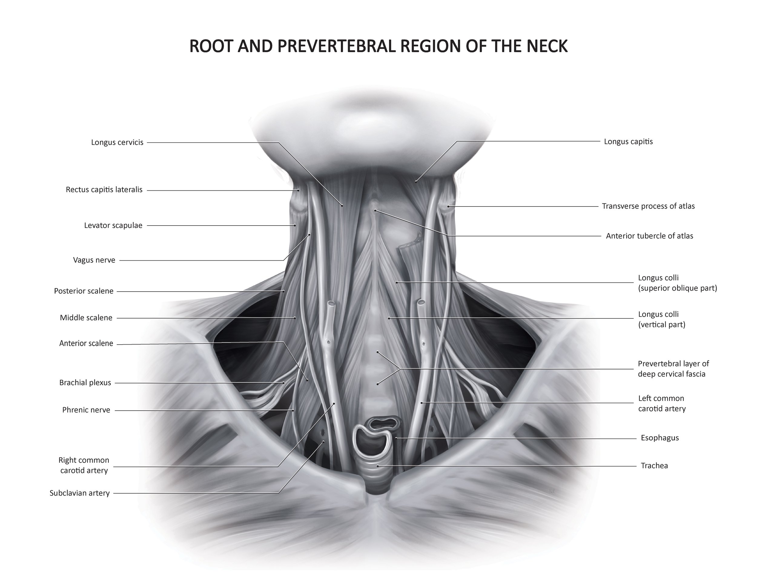
Prosection from Grant’s Museum
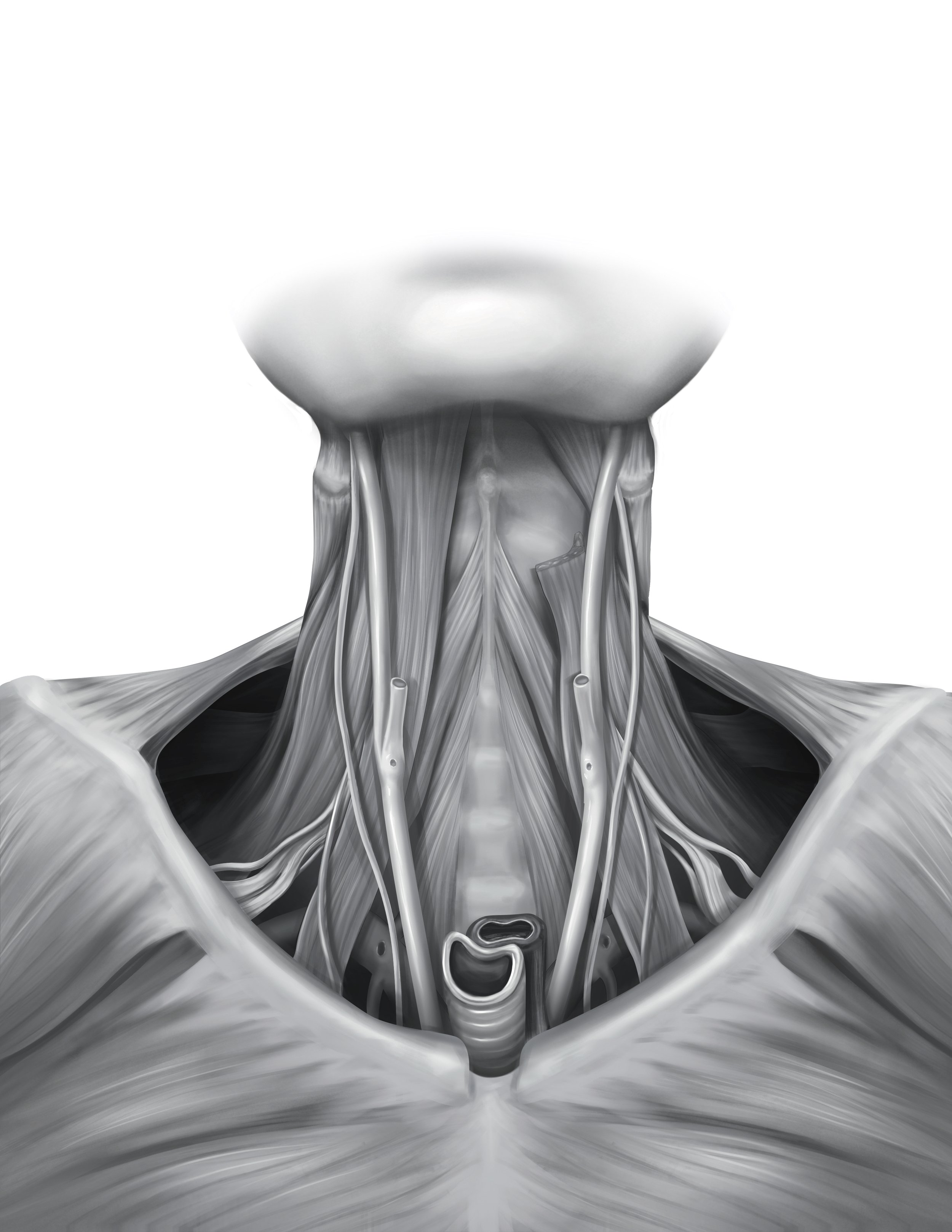
University of Toronto students have access to a rare resource: Grant’s Museum of Human Anatomy. Only medical students can access the room, which contains hundreds of prosected specimens from every part of the body. Photography is strictly prohibited.
This assignment asked students to pick one specimen and sketch it from life across several days of observation. I chose to portray the root and prevertebral region of the neck due to it’s intricacy and challenge. Final renderings were painted in grayscale using Photoshop.
